Key Takeaways
- MRI contrast injector safety depends on precise equipment design, adherence to ACR and FDA standards, and rigorous protocols around contrast media risks and extravasation.
- Shared responsibility among radiologists, technologists, and nurses—with clearly defined roles for screening, setup, monitoring, and documentation—is essential to prevent protocol gaps.
- A structured safety program, including written policies, checklists, verified consumables, and regular protocol reviews, reduces equipment failures, workflow disruption, and medicolegal exposure.
- Stepwise safety procedures covering room and injector preparation, patient screening, IV access verification, parameter double-checking, active monitoring, and post-injection assessment create layered protection.
- Robust emergency preparedness—early recognition of reactions, standardized response steps, coordination with emergency teams, and thorough documentation—turns rare adverse events into opportunities for continuous safety improvement.
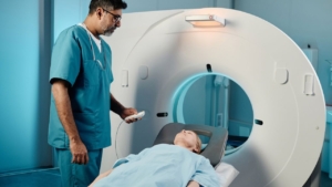
MRI contrast injectors sit at the intersection of advanced imaging technology and patient safety. These systems automate the delivery of gadolinium-based contrast agents in a high-field magnetic environment, where even small errors in setup, dosing, or monitoring can lead to extravasation, adverse reactions, workflow disruption, or regulatory scrutiny. Because injectors control both the volume and rate of contrast delivery, they are central to safeguarding patients, protecting staff, and preserving image quality.
For medical staff working in MRI, safety is not just about having the right device—it is about following the right protocols every time. From complying with ACR Manual on Contrast Media guidance and FDA-cleared indications, to using verified consumables, defining team roles, and preparing for emergencies, contrast injector safety requires a structured, team-based approach. This article explains how MRI contrast injectors work, what risks they carry, which standards apply, and, most importantly, what day-to-day safety protocols staff should follow to prevent accidents and ensure consistent patient protection.
What Is an MRI Contrast Injector and Why Is Safety So Critical?
MRI contrast injectors automate the delivery of gadolinium-based contrast agents during magnetic resonance imaging procedures. These specialized devices must function flawlessly in high-field magnetic environments while ensuring precise dosing, preventing extravasation, and protecting both patients and staff. MRI injector safety protocols form the foundation of reliable medical imaging safety procedures, as even minor equipment failures or protocol deviations can lead to adverse patient outcomes, workflow disruptions, and regulatory compliance issues.
How Do MRI Contrast Injectors Operate Within the MRI Environment?
MRI contrast injectors like the OptiStar® Elite use ultrasonic non-magnetic motors specifically engineered for safe operation up to 3 Tesla field strengths. The ceiling-mounted design eliminates floor cabling, reducing trip hazards and creating safer navigation paths for patients and staff in the scan room. These dual-head contrast delivery systems support complex injection protocols, including multi-phase sequences and variable flow rates tailored to specific diagnostic imaging requirements.
Modern MRI contrast administration protocols demand equipment that withstands intense magnetic fields without compromising injection precision or introducing ferromagnetic hazards. The non-magnetic architecture ensures consistent performance while maintaining the integrity of both the imaging environment and patient safety parameters.
What Types of MRI Contrast Media Are Used and What Risks Do They Carry?
Gadolinium-Based Contrast Agents (GBCAs) serve as the primary contrast media for MRI procedures. Clinical data spanning 25 years and 50 million doses show gadoterate meglumine carries a 0.3% adverse reaction rate, with most reactions manifesting as non-severe skin reactions, nausea, headache, or injection site pain affecting more than 2% of patients in clinical trials. These figures establish important benchmarks for contrast injector best practices and risk assessment.
MRI extravasation occurs at a rate of just 0.06%, with no serious complications typically reported. GBCM extravasation presents lower toxicity risks compared to iodinated contrast media due to smaller injected volumes and inherently reduced tissue reactivity, making MRI contrast administration protocols comparatively safer when proper techniques are followed.
Which Standards and Guidelines Govern Safe MRI Contrast Injector Use?
The ACR Manual on Contrast Media (2024) provides comprehensive, evidence-based guidelines that define industry standards for MRI injector safety and radiology staff training guidelines. This authoritative resource covers patient screening, injection techniques, adverse event management, and equipment protocols that form the regulatory backbone of safe practice.
FDA 510(k) clearance (K073592, cleared May 7, 2008) for devices like the OptiStar® Elite establishes baseline safety and efficacy standards for injecting MR contrast media and flushing solutions into the patient’s vascular system. Compliance with these federal regulations ensures equipment meets minimum performance thresholds and supports defensible clinical protocols.
Who Is Responsible for MRI Contrast Injector Safety Within the Imaging Team?
MRI injector safety requires shared accountability across multiple clinical roles, with radiologists providing medical oversight while technologists and nurses execute hands-on procedures. Establishing clear role definitions prevents gaps in patient monitoring, ensures consistent protocol adherence, and creates redundant safety checks throughout the contrast administration workflow. Effective medical imaging safety procedures depend on every team member understanding their specific responsibilities and maintaining open communication channels during each injection sequence.
How Do Radiologists, Technologists, and Nurses Share Safety Responsibilities?
According to ACR guidelines, radiologists, radiologic technologists, or nurses may administer contrast media, subject to state law requirements and institutional credentialing policies. Radiologists maintain oversight responsibility for contrast administration decisions, including patient screening determinations, dose selection, and management of adverse reactions requiring medical intervention.
This multi-disciplinary approach to contrast injector best practices distributes technical execution across trained personnel while preserving physician authority for clinical judgment calls. State regulations vary significantly, making institutional policies essential for defining which roles can independently operate injectors versus requiring direct supervision.
How Should Roles Be Defined for Screening, Setup, Monitoring, and Documentation?
All personnel involved in MRI contrast administration protocols must receive training in rapid recognition, assessment, diagnosis, and treatment of contrast reactions, with documented proficiency in basic life support. Clear role delineation should specify who conducts patient screening, who performs injector setup and quality checks, who maintains visual monitoring during injection, and who completes required documentation.
Overlapping responsibilities create redundancy that catches errors before they reach patients. One staff member might screen and establish IV access while another programs injection parameters, followed by independent verification before contrast delivery begins. This systematic approach to radiology staff training guidelines reduces single-point failures and reinforces accountability at each procedural step.
How Can Leadership Build a Strong Safety Culture Around MRI Contrast Injections?
Leadership cultivates a safety culture by investing in comprehensive training programs that align with ACR recommendations, such as Guerbet’s online courses covering fundamental MRI safety and device-specific instruction for systems like the OptiStar® Elite injector. Regular competency assessments ensure staff maintain current knowledge as protocols evolve and new equipment enters service.
Fostering open communication and non-punitive incident reporting transforms near-misses into learning opportunities rather than blame exercises. When staff feel safe reporting close calls or procedure deviations, departments gain actionable intelligence for improving MRI injector safety protocols before adverse events occur. This proactive stance strengthens both compliance and clinical outcomes.
How Can MRI Departments Structure a Comprehensive Contrast Injector Safety Program?
A comprehensive safety program integrates equipment policies, protocol maintenance schedules, and communication standards into a cohesive framework that prevents errors and streamlines workflows. MRI contrast administration protocols succeed when departments move beyond reactive troubleshooting to proactive risk mitigation through documented policies, regular audits, and systematic staff communication. Structured programs create consistency across shifts, reduce variability in patient outcomes, and provide clear accountability when incidents require review.
What Should an MRI Contrast Injector Safety Policy and Checklist Include?
Safety policies must mandate the use of verified consumables to ensure reliable operation and prevent equipment malfunctions that compromise patient safety. Verified consumables deliver three critical advantages: time and cost savings by preventing re-examinations, equipment downtime, and revenue loss from canceled procedures; reduced repair costs by avoiding expensive injector failures not covered by service agreements; and enhanced safety that directly protects patients and medical staff from preventable complications.
Policies should explicitly address generic consumables, which can invalidate warranties and service contracts while introducing unpredictable performance variables. Contrast injector best practices require procurement standards that prioritize manufacturer-verified components over cost-cutting alternatives that increase long-term risk exposure and financial liability.
How Often Should Protocols, SOPs, and Risk Assessments Be Reviewed and Updated?
Protocols require immediate review whenever the ACR Manual on Contrast Media receives updates—most recently the 2024 edition—to incorporate new evidence-based recommendations into active clinical practice. Regular review cycles ensure alignment with evolving industry standards, emerging safety data, and technological advancements in injection systems.
Annual or biannual safety audits provide structured opportunities to assess protocol effectiveness, identify gaps between written procedures and actual practice, and implement corrective actions. These scheduled reviews transform medical imaging safety procedures from static documents into living frameworks that adapt to operational realities and regulatory changes.
How Can Communication and Handoff Processes Reduce Injector-Related Errors?
Patient engagement protocols demonstrably reduce extravasation risk through clear communication before and during contrast injection. Patients instructed to immediately report any discomfort, pain, or swelling become active participants in their own safety monitoring, creating an additional layer of detection beyond staff observation.
Standardized handoff communication between screening staff, injector operators, and monitoring personnel prevents critical information loss during care transitions. Structured handoff tools—such as SBAR (Situation-Background-Assessment-Recommendation) formats—ensure consistent transfer of patient risk factors, IV access quality, programmed injection parameters, and monitoring requirements. This systematic approach to radiology staff training guidelines eliminates ambiguity and reduces reliance on assumptions or memory during high-volume clinical operations.
What Safety Protocols Should MRI Staff Follow to Prevent Contrast Injector Accidents and Ensure Patient Protection?
Preventing contrast injector accidents demands rigorous adherence to sequential safety protocols that address equipment preparation, patient screening, IV access verification, parameter checking, real-time monitoring, and post-injection assessment. These layered defenses create redundant safeguards that catch potential failures before they reach patients. Comprehensive MRI contrast administration protocols translate evidence-based guidelines into actionable steps that frontline staff execute during every injection sequence.
How Should MRI Staff Prepare the Room, Injector, and Equipment Before Starting the List?
Meticulous preparation of the power injection apparatus minimizes extravasation and air embolism that compromise patient outcomes. Standard procedures require staff to clear all air from syringes and pressure tubing, reorient syringes with tubing directed downward to facilitate air bubble migration, and position injector components to allow unobstructed table movement without creating tension on IV lines.
This preparatory phase establishes the foundation for safe contrast delivery by eliminating preventable equipment-related complications. MRI injector safety begins before the first patient enters the scan room, making equipment checks non-negotiable elements of daily workflow rather than optional quality measures.
How Should Patients Be Screened for MRI Safety and Contrast-Related Risk Factors?
Thorough screening for contraindications must identify patients with histories of severe allergic-like reactions or renal impairment that elevate adverse event risk. ACR Manual Chapter 4 provides detailed patient selection and preparation strategies that inform institutional screening protocols and decision algorithms.
A history of prior severe contrast reaction constitutes a relative contraindication for future administration of the same contrast medium class. These patients require alternative imaging strategies, premedication protocols, or heightened monitoring that standard workflows may not accommodate. Effective screening separates routine cases from high-risk scenarios requiring specialized management.
How Should IV Access Be Established, Verified, and Secured Before Injection?
Contrast injector best practices mandate flexible plastic cannulas rather than metal needles for power injection. A 20-gauge or larger catheter is preferred for flow rates at or exceeding 3 mL/sec, though 22-gauge catheters may tolerate up to 5 mL/sec. Antecubital or large forearm veins serve as preferred injection sites, while more peripheral locations like hands or wrists require reduced flow rates of 1-2 mL/sec to prevent vessel injury.
The Patency Check® feature on systems like the OptiStar® Elite confirms venous access before contrast administration through automated saline test injection. Staff should verify catheter location by checking for blood backflow—though absence doesn’t always indicate improper placement—and perform manual saline test flushes. Secure catheter fastening with appropriate devices prevents dislodgement during patient positioning or injection pressure fluctuations.
How Should Contrast Dose and Injector Parameters Be Checked and Double-Verified?
Modern injectors like the OptiStar® Elite offer volume precision down to 0.1 mL, enabling accurate delivery of lower GBCA doses tailored to patient-specific imaging requirements. The Timing Bolus® feature ensures precise synchronization between contrast injection and pulse sequence acquisition, optimizing enhancement while minimizing waste.
Staff must consult the FDA package inserts for appropriate doses and concentrations before programming injection parameters. Automated injections and multiple-phase protocols standardize delivery sequences, reducing human error inherent in manual processes. Medical imaging safety procedures require independent double-verification of programmed parameters before initiating injection, creating a final checkpoint that catches entry errors or inappropriate protocol selection.
How Should MRI Staff Monitor the Patient and Injector During Contrast Delivery?
Automatic Pressure Control manages injection pressure dynamically, reducing vein rupture risk and high-pressure complications that occur when resistance exceeds safe thresholds. Variable Drip Mode provides flexibility in contrast delivery, allowing operators to adjust flow characteristics based on real-time patient needs or unexpected vascular responses.
Injection must be discontinued immediately if patients report pain or swelling at the injection site, as these symptoms often precede clinically significant extravasation. Continuous visual monitoring of both the injection site and overall patient status throughout delivery enables rapid detection of complications before they progress to serious injury. This active surveillance transforms passive observation into purposeful safety monitoring.
What Post-Injection Checks, Observation, and Documentation Steps Are Essential?
Patients experiencing mild allergic-like reactions require observation for 20-30 minutes to ensure symptoms resolve without progression. Moderate to severe reactions demand prompt and aggressive intervention following established emergency protocols that radiology staff training guidelines should cover in depth.
Any patient with suspected or confirmed extravasation must undergo examination for tenderness, swelling, erythema, paresthesia, range of motion, and perfusion status. Complete documentation of injection parameters, patient responses, and any adverse events creates medicolegal records and quality improvement data that inform future protocol refinements. Systematic documentation closes the loop on comprehensive safety programs by capturing outcomes that validate or challenge existing practices.
How Should MRI Staff Manage Emergencies and Adverse Reactions Related to Contrast Injections?
Emergency preparedness separates competent imaging departments from those unprepared for life-threatening complications. While adverse events during MRI contrast injections remain statistically rare, staff must recognize warning signs immediately and execute standardized response protocols without hesitation. Effective radiology staff training guidelines ensure every team member can initiate appropriate interventions while coordinating with broader emergency response systems when complications exceed departmental capabilities.
How Can Teams Recognize Early Signs of Extravasation or Contrast Reactions?
MRI extravasation occurs at a benchmark incidence of 0.06%, making it uncommon but not negligible in high-volume practices. High-risk patient factors include uncommunicative patients unable to report symptoms, altered circulation in the injected extremity, prior radiation exposure to the injection site, and peripheral injection locations such as hands, feet, or ankles, where vessel fragility increases complication severity.
More viscous contrast materials elevate extravasation risk through increased mechanical stress on vessel walls during power injection. Early signs demand immediate attention: patient-reported pain or burning sensation at the injection site, visible swelling indicating tissue infiltration, and resistance feedback from injector pressure monitoring systems. Recognizing these indicators within seconds of onset prevents minor leaks from progressing to compartment syndrome or tissue necrosis.
What Immediate Steps Should Staff Take When an Adverse Event Occurs?
The immediate assessment protocol follows a systematic 5-point check: Airway patency, Breathing adequacy, Circulation status, Disability assessment of neurologic function, and Exposure for full body evaluation. This structured approach prevents tunnel vision on the injection site while missing systemic reactions requiring urgent intervention.
For confirmed extravasation, elevate the affected extremity and apply cold or warm compresses according to contrast media type and institutional protocol. Patients with documented histories of severe reactions may benefit from premedication with corticosteroids administered 4-5 hours before injection, with optional diphenhydramine for additional prophylaxis. Discontinue injection immediately upon adverse event recognition while maintaining IV access for emergency medication administration. These contrast injector best practices balance stopping ongoing injury against preserving vascular access critical for resuscitation drugs.
How Should MRI Teams Coordinate With Emergency Response and Document the Event?
Patients discharged after extravasation require clear written instructions detailing symptoms that demand immediate medical attention: worsening pain, paresthesia indicating nerve involvement, or skin ulceration suggesting tissue breakdown. Verbal instructions alone prove insufficient, given patient stress and information overload during adverse events.
Surgical consultation becomes warranted for severe extravasation injuries manifesting as severe or progressive pain, decreased capillary refill indicating vascular compromise, altered sensation, worsening range of motion, or visible skin ulceration and blistering. Complete incident documentation must capture volume extravasated, time course from injection start to recognition, all interventions performed, patient education provided, and follow-up arrangements made. This comprehensive record serves quality improvement analysis, medicolegal protection, and regulatory reporting requirements that govern medical imaging safety procedures. Thorough documentation transforms isolated incidents into learning opportunities that strengthen institutional MRI injector safety protocols for future patients.
Raise Your MRI Injector Safety Standards With Hitech Global Medical Services Co.
Safe MRI contrast injection does not happen by chance—it comes from clear policies, reliable equipment, disciplined workflows, and a team that trains and practices together. When you combine evidence-based guidelines with practical checklists and strong communication, you reduce complications, protect patients, and give your staff confidence in every injection.
At Hitech Global Medical Services Co., we help you turn these best practices into everyday reality. We work with your team to review current injector workflows, align policies with ACR guidance, choose verified consumables, and design training that prepares staff for both routine lists and rare emergencies. If you are ready to strengthen MRI contrast injector safety, reduce risk, and support your staff with better tools and protocols, we invite you to connect with us. Together, we can build a safer, more reliable MRI environment for every patient you scan.